Click Here To Download Fundamentals of Nanoscience Unit V Notes as PDF
Showing posts with label Fundamentals of Nano Science. Show all posts
Showing posts with label Fundamentals of Nano Science. Show all posts
Nanosensors in Optics - V Unit Notes
Nanosensors in Optics
Nanosensors are devices with
dimensions on the nanometer scale that are capable of monitoring the presence
of a specific chemical or class of chemicals. Nanosensors which employ optical transduction methods are called optical Nanosensors.
Optical nanosensors can
generally be classified into one of two different classes: 1) chemical
nanosensors, or 2) nanobiosensors, depending on the type of recognition element
(chemical or biochemical) used to provide specificity to the sensor. Small
sizes of these sensors allow them to be inserted and precisely positioned
within individual cells to obtain spatially localized measurements of chemical
species in real time.
Fiber optic nanosensors employ
fiber optics that have been tapered on one end to diameters typically ranging
between 20 and 100 nm. Excitons or evanescent fields continue to travel through
the remainder of the tapered fiber’s tip, providing the necessary excitation
energy. Excitation using such a sensor is highly localized, allowing only
species close to the fiber’s tip to be excited.
The most significant
applications of fiber optic nanoprobes to NSOM analyses of biological samples occurred
when a single dye-labeled DNA molecule was detected using near-field
surface-enhanced resonance Raman spectroscopy (NFSERRS). In that work, dye-labeled
DNA strands were spotted onto a surface-enhanced Raman spectroscopy (SERS) substrate
that was prepared by evaporating silver on a nanoparticle-coated surface. Following
preparation of the sample, a fiber optic nanoprobe was raster-scanned over the
sample’s surface, illuminating it point by point, while the resulting Raman
signals were measured with a charge-coupled device (CCD). Based on the
intensity of the Raman signals measured at every location, a two-dimensional
image of the DNA molecules was reconstructed and normalized
for surface topography based on the intensity of the Rayleigh scatter.
for surface topography based on the intensity of the Rayleigh scatter.
FIBER OPTIC CHEMICAL NANOSENSORS
Fiber
optic chemical nanosensors have chemical recognition elements (e.g.,
fluorescent indicator dyes, etc.) bound to the tapered tip of the fiber to
provide a degree of specificity. It is important to employ a sensitive detection
system, such as the one shown in the following figure.
In
such a system, the sample is excited by launching an intense light source
(e.g., laser) into the proximal end of the fiber optic nanosensor. The
nanosensor is then positioned in the desired location using an x–y–z micromanipulator
or piezoelectric positioning system mounted on a microscope. Once in place, the
fluorescent indicator dye immobilized on the tip of the fiber is excited, and
the resulting fluorescence emission is collected and filtered by the microscope
before being detected with either a photomultiplier tube (PMT) or a CCD.
One
advance in the last several years has been the development of
nanoparticle-based optochemical sensors, with nanometer-scale sizes in all
three dimensions. Because of the small sizes of these sensors, a large number
of them can be implanted within an individual cell at one time, allowing for
the monitoring of many locations simultaneously. Although many different
nanoparticle-based sensors are currently being developed, three main classes
have already shown a great deal of promise for intracellular analyses. These
three classes are
·
Quantum
dot-based nanobiosensors
·
Polymer-encapsulated
nanosensors known as PEBBLEs
·
Phospholipid-based
nanosensors
Nanosensors in Biomedical Field - V Unit Notes
Nanosensors in
Biomedical Field
Biosensors are sensors for
detecting biological entities such as proteins, drugs, specific viruses, cancer
cells etc. In vivo detection of these happens in variety of ways naturally. For
an example, when a body is first exposed to an allergen, the body creates
antibodies that will recognize that allergen if it appear again in the body.
This triggers allergy response system the body to release histamine. Glucose detection
is important in biosensing. Type I diabetics has to monitor their blood sugar
levels continuously. Nanoscale structures may advance it in a big way.
DNA sensing is another important
area in which nanosensing can play a potential role. Using the ability of DNA
to bind to a complementary strand and not to bind to anything else presence of any microorganism with known DNA sequence. For
instance, to sense the structure with the sequence CGCTTC a complementary strand
GCGCAAG can be used.
A single strand of, say, six
bases can contain 4,096 different combinations. Consequently, if a particular
biological target such as botulism or strep or scarlet fever has a known DNA
sequence, it is possible to target a short section of that DNA sequence that
can be uniquely sensed, without any errors, by an appropriate single-strand
complementary structure.
This is called DNA finger printing.
This is called DNA finger printing.
Generalization of this method
leads to lab-on-a-chip concept. Microlaboratories capable of sensing viral and
bacterial diseases are possible with this technology. Finally, biochips could
be used to sense either particular DNA signatures or particular protein
signatures known to be defects that can result in disease.
One of the great challenges of DNA
sensing is to amplify the effects of hybridization so that they can be easily
measured. One way to provide this amplification is to change the optical
properties of gold or silver nanodots that are attached to the DNA. Chad Mirkin,
Robert Letsinger, and their groups at Northwestern pioneered the combination of
quantum optical effects and molecular recognition (complementary DNA binding).
Their scheme and some actual results are shown in the following figure.
 |
| DNA Viral Detection |
The upper schematic shows how the
nanodots in a colorimetric sensor are brought together upon binding to the DNA
target (in this case anthrax). The clustered dots have a different color than
the unclustered ones as is shown in the photograph below them.
By exposing the single strands
of DNA that are attached to the gold nanodots, the sensor recognizes the target strands of DNA, which causes the gold nanospheres
to come closer together and, as in those recurring stained glass windows, change
color.
In a protein biosensor a molecular
nanostructure containing a biological binding site is attached to the gold
nanoparticles. The binding site is designed to recognize a particular protein analyte. When that analyte appears in solution,
it binds to the recognition site, which changes the chemical and physical environment of the
gold dot, whose color is then slightly changed. This change can be measured.
In
the electronic nose, a random polymer, or mix of polymers, is spread between
electrodes. When the molecules to be smelled land on the polymer(s), the
conductivity properties in particular regions will change in a particular way
that is specific to any given analyte.
Nanomaterials Applications in Electronics - V Unit Notes
· Transistors
have gotten smaller through nanotechnology. Around 2001, a typical transistor
was 130 to 250 nanometers in size. In 2014, Intel created a 14 nanometer
transistor, then IBM created the first seven nanometer transistor in 2015, and
then Lawrence Berkeley National Lab demonstrated a one nanometer transistor in
2016. Research is on going to use single molecules as transistors.
· Using
magnetic random access memory (MRAM), computers will be able to “boot” almost
instantly. MRAM is enabled by nanometer‐scale magnetic tunnel junctions and can
quickly and effectively save data during a system shutdown or enable
resume‐play features.
· Ultra-high
definition displays and televisions are now being sold that use quantum dots to
produce more vibrant colors, while being energy efficient.
· Flexible,
bendable, foldable, rollable and stretchable electronics are reaching into
various sectors and are being integrated into a variety of products,
including wearables, medical applications, aerospace applications,
and the Internet of Things. Flexible electronics have been developed using, for
example, semiconductor nanomembranes for applications in smartphone and
e-reader displays. Nanomaterials like graphene and cellulosic nanomaterials are
being used for various types of flexible electronics to enable wearable and
“tattoo” sensors, photovoltaics that can be sewn onto clothing, and electronic
paper that can be rolled up. Making flat, flexible, lightweight, non-brittle,
highly efficient electronics opens the door to countless smart products.
· Computing
and electronic products include Flash memory chips for smart
phones and thumb drives; ultra-responsive hearing aids;
antimicrobial/antibacterial coatings on keyboards and cell phone casings;
conductive inks for printed electronics for RFID/smart cards/smart packaging;
and flexible displays for e-book readers.
· Nanoparticle
copper suspensions have been developed as a safer, cheaper, and more reliable
alternative to lead-based solder and other hazardous materials commonly used to
fuse electronics in the assembly process.
· DNA
can be used as scaffolding for assembling molecules into electronic circuitry.
This is used to integrate novel devices at densities far beyond those possible
lithographic techniques. Studies on DNA showed a wide range of electron
transport behavior. DNA can act as an insulator, a semiconductor, a conductor
and a super conductor offering future bio-compatible devices and circuits.
· Nanomaterials
research, especially molecular electronics has demonstrated the new electrical
element ‘Memristor’. Memristor has the capability of remembering the current it
experienced in the past. So Memristor
research offers a possibility to implement 50Gbytes of low-power memory in
mobile devices.
· Nanomagnetics
provide an opportunity to realize zero-energy switching logic gates and memory.
Phase change memory (PCM) is considered one of the most promising candidates
for next-generation nonvolatile memory, based on its excellent characteristics
of high speed, large sense margin, good endurance, and high scalability.
Nano Materials applications in Magnetism - V Unit Notes
Nano Materials applications in Magnetism
Cancer Treatment
Magnetic
nanoparticles have been examined for use in an experimental cancer
treatment called magnetic hyperthermia in which an
alternating magnetic field (AMF) is used to heat the nanoparticles. To achieve
sufficient magnetic nanoparticle heating, the AMF typically has a frequency
between 100–500 kHz, although significant research has been done at lower
frequencies as well as frequencies as high as 10 MHz, with the amplitude
of the field usually between 8-16kAm−1
Nano Particle Applications - V Unit Notes
Nano Particle Applications
A few of the applications of Nano particles are listed out
below.
- Nano particles are used in sunscreen and skin cream as optical filters
- Dirt repellents for cars and windows
- Flat screens
- Single electron transistors
- Nanowheels, nanogears, nanofilters
- Drug pumps located in the human body, long-term depots
- Luminescent devices
- Energy storage (hydrogen in zeolites)
- Electronic devices
Applications of Fullerene Nano Particles
- Particle absorption filters for cigarettes
- Chromatography
- Molecular containers
- Sensor cover layers for surface wave devices
- Additives in fuels
- Lubricants
- Catalysts for hydrogenation
- Photocatalysts for the production of atomic oxygen in laser therapy
- Production of artificial diamonds
- Functional polymers, photoconductive films
- Alkali metal MC60 chain formation (linear conductivity)
- Superconductivity (doping with alkali metals)
- Ion engines
- Raw material for AIDS drugs
- Tools that are harder than diamond
- Nanoelectronic devices
Nanolayer Applications - V Unit Notes
Nanolayer Applications
Nanolayers
have found widespread applications in recent times. A few of the applications
are listed below.
- MOS(Metal Oxide Semiconductor) gate oxide
- Field oxide
- SOI (Silicon On Insulator)
- Amorphous layers for heterojunction solar cells, Thin Film Transistors and optical sensors
- MOS channels
- Counter-doped Si layers for p-n junctions and transistors
- Recrystallized layers on dielectrics for device production
- Oxide as implantation and diffusion masks
- Oxide for the photolithography
- Silicides or metals
- Epitaxial layers for transistors, laser, quantum detectors
- ITO for anti-reflection and charge collection in solar cells
- Back side surface field (BSF) layers in solar cells
- Metal layers for glasses, lenses, beam splitters, interferometers
- Anti-corrosion and passive layers
Scanning Tunneling Microscope - Links for Netizens and Additional Material
- A Wikipedia Schematics, If you find this more appropriate and to your liking use this:
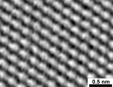
- Atom Level Resolution of Carbon atoms on a graphite surface. Let Boltzman Soul rest in Peace!

- Gold Atoms, They are not yellow of course, a suitable false color applied by the computer.
| The Quantum Coral - The Most Famous Image Produced by STM |
- See this animation video
- This is a 3 in 1 video about STM,SEM and AFM, which we shall see in future lectures.
- World's Smallest Animation Movie - Just check this animation "A Boy and His Atom" by IBM.
- And How They Moved These O-Atoms ! அணுவைத் துளைத்து ஏழ் கடலைப் புகட்டிக் குறுகத் தறித்த IBM !
- Gerd Binnig and Heinrich Rohrer were awarded Nobel Prize in 1986 for their invention of STM.
Syllabus for Fundamentals of Nanoscience
III YEAR – VI
SEMESTER
COURSE CODE:
4BPHE3C
ELECTIVE
COURSE III (C) – FUNDAMENTALS OF
NANOSCIENCE
Unit I Introduction
Introduction to
Nanotechnology – Background and definition of Nanotechnology – Nano materials –
Size Dependence.
Types: Nanowires, Nanotubes,
Quantum Dots, Nanocomposites – Properties – Ideas about Nano materials
synthesis.
Unit II Carbon
Nano Tubes (CNT)
Introduction to
CNT – SWNT – MWNT – Properties. CNT based Nano
objects- Applications.
Unit III Fabrication
Fabrication methods – Top down
processes – Milling, lithographics, Machining process. Bottom–Up process – MBE
and MOVPE, liquid phase methods, colloidal and sol – gel methods – Self
Assembly
Unit IV Characterization
Scanning Probe
Microscopy – Principle of operation – Instrumentation – Scanning Tunneling
Microscopy – STM probe construction and measurement.
Atomic Force
Microscopy – Instrumentation and Analysis – Tunneling Electron Microscopy–
operation and measurement
Unit V Nano devices
and Applications
Optical memories, Nano materials
applications in magnetism – in electronics. Sensors – in Biomedical field – in
optics – Nano layer applications – Nano particle applications
Reference
1.
Hand book of Nanotechnology – Bharat Bhushan.
2.
Nano technology and Nano electronics – W. R. Fahrner
(Editor).
3.
Materials Science – P. Mani, G. Ranganath, R. N.
Jayaprakash.
4.
Nanotechnology – Mark Ratner, Daniel Ratner.
♣♣♣♣♣♣♣♣♣♣
Subscribe to:
Posts (Atom)


